Textbook:
MCQ:
Learning Outcomes:
Basics
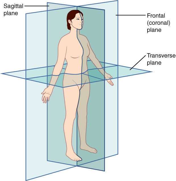
OpenStax College. Anatomy & Physiology, Connexions Web site. http://cnx.org/content/col11496/1.6/, Jun 19, 2013.
Anatomical terms of relationship:
Basics (should know): superior, inferior, posterior, anterior, medial, lateral
Others:
- caudal = towards feet (or tail)
- rostral = towards rostrum('beak' in latin), anterior
- palmar surface of hand= volar surface
- dorsum of hand = back of hand, opposite palm
- plantar surface of foot = sole
- dorsum of foot = top of foot
Terms of movement
Basics (should know): flexion (reducing angle), extension (increasing angle), elevation, depression, abduction (away from middle), adduction (towards middle)
Others:
- opposition of thumb = pad of 1st digit brought to another digit
- eversion = foot away from median plane VS inversion = foot towards median
- protrusion = movement anteriorly VS retrusion = movement posteriorly
- supination = palms up VS pronation = palms down
Bursae and potential spaces
Bursa: a closed sac lined with synovial membrane and containing a thin film of synovial fluid. It reduces friction where skin, tendon, or muscle moves over bone.
- Anatomical (constant) bursae form in utero, e.g. subacromial and prepatellar.
- Adventitious bursae form after birth in response to friction, e.g. over a bunion.
- Communicating bursae connect with a joint cavity.
Bursitis can be caused by trauma, repetitive friction, crystals, infection, inflammatory arthritis. Septic bursitis mostly affects superficial bursae (olecranon, pre-patellar) and organism (80% Staph aureus) enter through breaks in the skin. A septic aspirate will show high WCC, neutrophils, positive gram stain, low glucose (note crystals don't exclude infection).
Upper Limb:
Subacromical(subdeltoid) bursa: between acromion/deltoid/coracoacromial arch and supraspinatus tendon. Fluid in the bursa can suggest a cuff tear.
Olecranon bursa: subcuaneous over the olecranon. Septic bursitis (staph aureus), traumatic, or "students elbow"
Ulnar and Radial bursae (tendon sheaths): the little finger flexor (palmar surface) sheath is continuous with the ulnar bursa (the common flexor sheath around FDS and FDP in the carpal tunnel). the thumb (FPL) sheath is continuous with the radial bursa. The radial and ulnar bursae often communicate allowing a "horseshoe abscess" where infection spreads from the thumb to the middle finger. Flexor tenosynovitis (inflammation of flexor sheath) can present as a finger in flexed resting position, tenderness along the sheath and pain on passive extension of the finger (Kanavels signs).
Lower limb:
Prepatellar bursa: subcutaneous, anterior to the patella. "Housemaids knee" (kneeling forward), septic.
Superficial Infrapatellar bursa: subcutaneous, over the tibial tuberosity/patellar ligament. "Clergymans knee" (kneeling upright)
Deep infrapatellar bursa: between patellar ligament and tibia, behind infrapatellar fat pat. Not usually palpable.
Suprapatelllar bursa: deep to quadriceps muscle, above the patella. Articularis genus holds it up. Communicating with knee joint, so knee effusions fill it. Common site for knee aspiration.
Semimembranosis-gastrocnemius bursa: popliteal fossa, communicating with knee joint through a valve-like opening. Bakers cyst.
Pes anserine bursa: between sartorius/gracilis/semitendinosis insertion and the medial tibia. Common cause of medial knee pain in obesity and OA.
Trochanteric bursa: between gluteus maximus/ITB and the greater trochanter. Cause of lateral hip pain.
Iliopsoas bursa: deep to psoas, anterior to hip capsule, communicating in 15% of patients with joint. Largest synovial bursa. Hip septic arthritis can track into it
Ischial bursa: over ischial tiberosity. "Weavers bottom" (prolonged sitting)
Retrocalcaneal bursa: between achilles tendon and calcaneous. Haglunds deformity.
Subcutaneous calcaneal bursa: between skin and achilles tendon. "Pump bump" from shoe friction.
Potential spaces
Head and neck
Cranial epidural: potential space. Site of extradural haematoma.
Subdural: potential space. Site of subdural haematoma.
VS subarachnoid and spinal epidural which are REAL spaces (not potential)
Retropharyngeal: from base of skill to T2, between the buccopharyngeal fascia (visceral layer of the pretracheal fascia) and the alar fascia. Retropharyngeal abscess more common in children under 5yrs.
Thoracic:
Pleural: normally 0.1-0.3mL/kg, roughly 10-20mL per side. Costo-diaphragmatic recess is the most dependent part.
Safe triangle for chest drains: anterior border of latissimus dorsi, lateral border of petoralis major, horizontal line at the 5th intercostal space, apex below the axilla. The neuromuscular bundle passes above the rib below and runs in the costal groove in the order VAN (vein, artery, nerve), from superior to inferior.
Pericardial cavity: normally 15-50mL. Outer later= Fibrous pericardium (inelastic) is attached to the central tendon of the diaphragm and continuous with the great vessel adventia. Inner layer = serous pericardium, which has parietal(outer sub-layer) and visceral (inner sub-layer) ayers. Rate of accumulation matters MORE than volume (i.e. 100-200mL acutely can cause tamponade).
Abdomen
Peritoneal cavity: normal fluid less than 50mL
Hepatorenal pouch (Morison’s pouch) is the most dependent upper abdominal space when supine. It is the first FAST window.
Rectovesical pouch in males and the rectouterine pouch (of Douglas) in females are the most dependent sites when upright.
Subphrenic spaces are classic sites for post-operative abscesses.
Paracolic gutters are pathways for spread. The right gutter connects the pelvis and the upper abdomen. The left is limited superiorly by the phrenicocolic ligament.
Perineum:
Superficial perineal pouch: bounded by Colles’ fascia, which is continuous with Scarpa’s fascia of the abdominal wall and dartos. In bulbar urethral rupture, urine extravasates into the scrotum, penis and lower anterior abdominal wall.
Bursae and potential spaces (especially in upper limbs)
Fascial compartments
Types of cartilage with examples
Types of joints with examples
Bone formation and markings, axial/appendicular skeletal components
Nervous system and its divisions: central/peripheral, somatic, autonomic, ganglia and neurotransmitters
Muscular system: skeletal, cardiac, smooth
What is a functional unit of muscle?
Vascular circuits and vessels
Lymphoid system and its functions/communications
Integumentary system: tension lines, laters and functions
Common anatomical variations
Veins vary the most and nerves the least
Hiltons law
Create Your Own Website With Webador